Even with excellent nutrition, cow comfort and management, even the best dairies deal with lameness to some degree, often occurring as a lesion in the rear outside claw.

“For dairy cattle housed on concrete flooring surfaces, it is not uncommon to observe that 90 percent of lameness is in the foot … 90 percent of that involves the rear feet … of that, nearly 90 percent involves the outside claw,” Dr. Jan Shearer, professor and dairy veterinary extension specialist at Iowa State University, says.
Sole ulcers and white-line disease are two of the most common lameness-causing claw disorders in cattle, according to Shearer. However, by applying proper therapy for these conditions and understanding their underlying causes, we can provide prompt relief and reduce the potential for future problems.
What causes claw lesions?
Shearer explains that sole ulcers are caused by a combination of mechanical and metabolic conditions.
Mechanical factors
Hard flooring surfaces can lead to horn overgrowth and unbalanced weight bearing within and between claws. In rear feet, the outer claw of the rear foot bears more weight; this leads to the acceleration of horn formation and an increase in sole thickness.
The cow compensates for this physical or “mechanical” overloading of her outer claw by rotating her feet outward (i.e., standing in a cow-hocked posture). This posture places more weight on the inner claw, which makes the cow more comfortable for a while, but eventually this imbalance in weight bearing leads to problems.
Metabolic factors
- Laminitis: Laminitis disrupts blood flow to the corium, causing inflammation and damage to tissues that suspend the third phalanx bone (P3) inside of the claw horn capsule. Because the corium produces hoof horn, this sort of disruption affects the quality of horn, resulting in weaker horn that is more prone to bruising and injury. In severe chronic cases, the hoof wall may flare, giving it a paddle-like appearance.
-
Hormonal changes: Occurring near the time of calving, hormones cause a weakening of the suspensory apparatus of P3. The result is sinking and rotation of P3 as the collagen fiber bundles (suspensory apparatus) suspending it are weakened.
It is believed that estrogen and relaxin may have a similar effect here as they do in the relaxation of the pelvic muscles and ligaments around the time of calving. Meanwhile, the cow is spending more time on her feet. These impacts are amplified if she is standing on a hard concrete surface, triggering further inflammation of the weakened tissue inside of the foot.
- Enzyme activity: Research has found that an enzyme known as “hoofase” has the potential to activate potent metalloproteinase enzymes capable of weakening the tissues supporting P3. These effects appear to be most significant during the transition period two weeks before calving through the first several weeks postpartum.
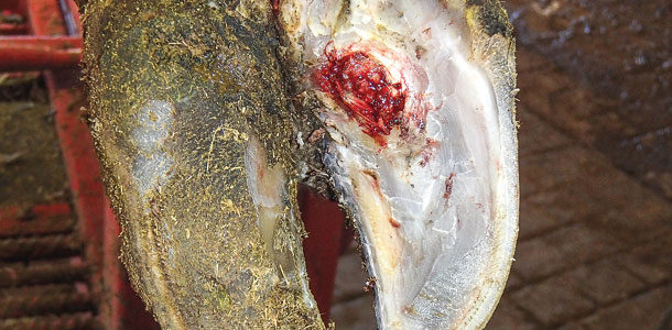
The resulting effect of these mechanical and metabolic factors is compression of the corium and the development of a sole ulcer. Sole ulcers are one of the most common of claw lesions, particularly in confinement facilities, according to Shearer. While the sole is the most common location, ulcers can also appear in the toe or heel.
Shearer notes that, according to research from Cornell University, the prevalence of sole ulcers and white-line disease is significantly associated with the cow’s body condition and thickness of the digital cushion. The digital cushion is a fat pad that absorbs the shock of the bones inside the claw and protects the corium.
If the cow is in poor body condition and the digital cushion is too thin, the sinking of P3 can damage the corium, resulting in a sole or heel ulcer. Conversely, cows with higher body condition scores are likely to have a thicker digital cushion and less potential for the development of claw lesions.
These factors combined plead the case for cow comfort, according to Shearer. Promote lying time with comfortable, clean stalls to minimize standing time, particularly during the transition period. Also, minimize standing time, especially on hard surfaces. Provide soft surfaces during the dry period, especially for first-calf heifers.
Treatment
Shearer says the objective for treating claw lesions is to “first, do no harm.” Because these ailments are not infectious, treatment beyond corrective trimming and a block to relieve weight bearing may be counterproductive.
- Trim: When trimming, he recommends balancing the load bearing between the weight-bearing surfaces of each claw. When corrective trimming is necessary, remove only the unhealthy horn without damaging adjacent healthy tissue. Removal of all loose and undermined horn creates an aerobic (oxygen-rich) environment that the wound needs for healing.
 “The less damage you do to adjacent healthy tissues, the quicker the cow recovers and less pain she feels later,” he adds.
-
Treat: According to a recent survey, most hoof trimmers and veterinarians apply blocks to the healthy claw in order to relieve weight bearing and alleviate the pain associated with the lesion on the affected claw.
Research demonstrates an “advantage as far as comfort for the cow” and improved lameness scores following block application three days post-treatment. Though there was less of a difference after seven days, providing weight-bearing relief during early recovery is an important benefit.
However, Shearer cautions, “Be careful not to be too aggressive with your therapy.”
The use of topical medications and wraps may cause irritation and even delay healing of lesions. Bandages or wraps should be reserved to control hemorrhages or protect delicate corium tissues exposed during corrective trimming. Bandages become heavily contaminated; therefore, it is best to remove or change them every day or two. This may not be practical in some herds.
- Recovery: Moving the cow to a special-needs pen or pasture for recuperation and monitoring during the recovery phase can provide a more comfortable environment with better footing. Be sure to re-check lesions to assess progress toward recovery and apply a new block if necessary.
If possible, avoid running these cows through footbaths with harsh, irritating chemicals while their wounds are healing.
Maintenance and preventative trimming should be done to remove abnormal overgrowth and balance weight bearing within and between claws. PD
Dr. Jan Shearer presented during the Wisconsin Hooftrimmer’s Getaway Weekend held in February.
PHOTO 1: Metabolic and mechanical factors contribute to the formation of sole ulcers, which are the most common claw lesion seen in confinement facilities.
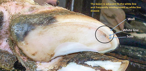
PHOTO 2: A thin sole toe ulcer has a similar appearance to white line disease, but it is caused by abrasive flooring, excessive wear or overtrimming.
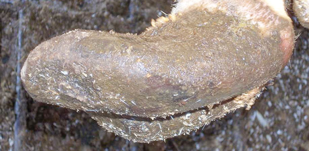
PHOTO 3: Severe, chronic cases of laminitis cause the hoof wall to flare, giving it a paddle-like appearance. Photos courtesy of Dr. Jan Shearer.
This article originally appeared in print with Is it a sole ulcer or white-line disease?

-
Peggy Coffeen
- Editor
- Progressive Dairyman
- Email Peggy Coffeen